
Dr.PARMITA DUTTA
Dr. (Mrs.) BARUAH MAMONI, Dr.Rajiv Kumar Das
Abstract
Acoustic neuromas account for 8% of intracranial tumours.Ophthalmic symptoms:16% cases.Papilledema: 8% cases.
A 40 year old female came with sudden onset diminution of vision R/E for 10 days, associated with holocranial headache, gradually progressive in nature since 2 months and right sided decreased hearing. V/A R/E:HMCF, V/A L/E:6/12, nystagmus present on looking towards right side.
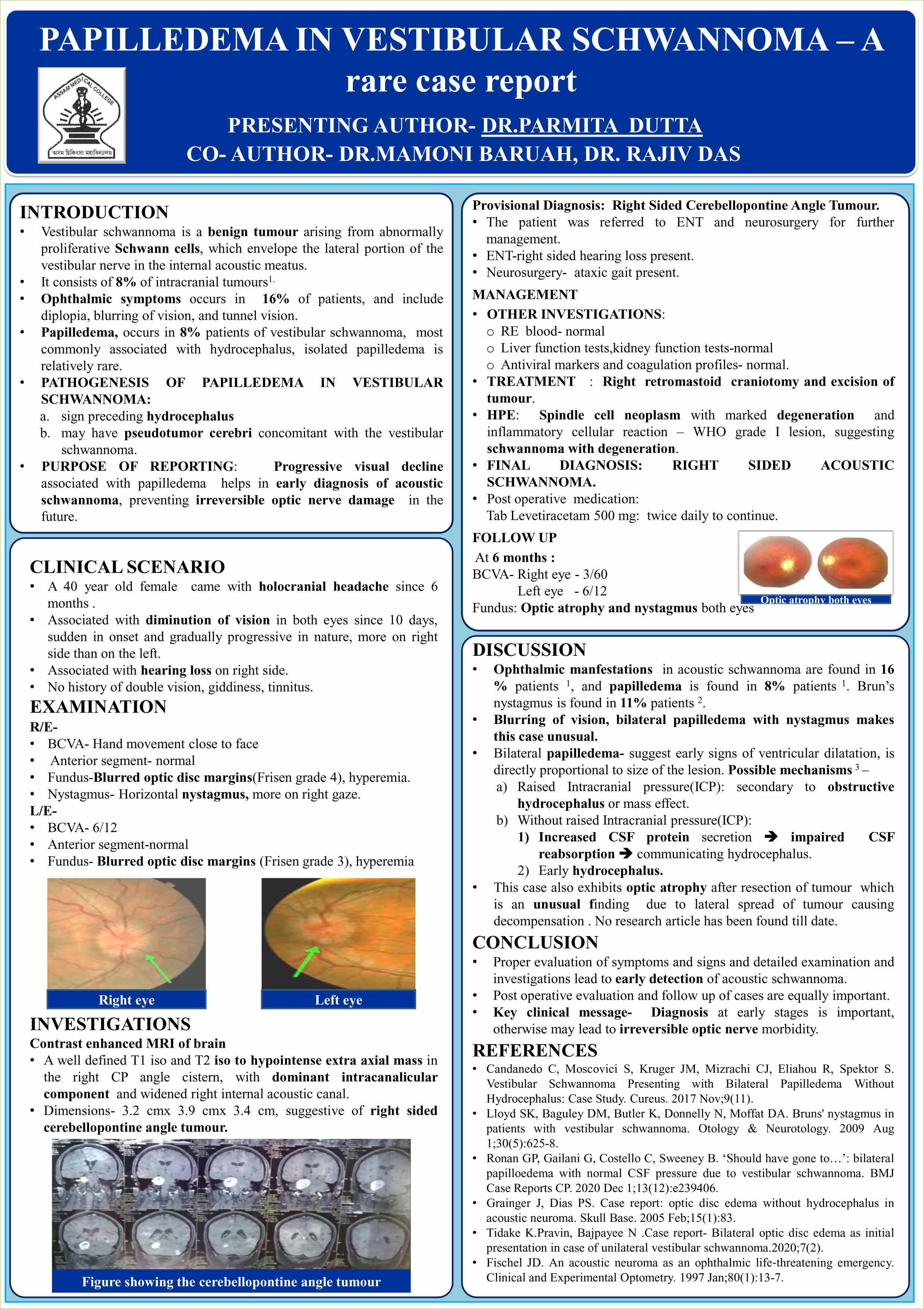
Fundus: papilledema (frisen grade 3) with dull foveal reflex B/E.
MRI-A well defined T1 isointense and T2 hypointense extra axial mass in right CP angle cistern,3.2cm(TR)x 3.9 cm(AP)x 3.4 cm, suggesting right acoustic schwannoma.
TREATMENT:Retromastoid craniotomy with surgical excision of the tumour.
HPE: degenerations, areas of proliferating spindle cells ,enlarged spindled hyperchromatic nuclei in sheets,suggesting spindle cell neoplasm with marked degeneration.
At 6 months follow up: V/A R/E:3/60,L/E:6/12, nystagmus present
fundus : optic atrophy B/E
There is no episode of recurrence till now.
Leave a Comment