
Dr. Madan Joshi
Dr.Apoorva A G, Dr. A.S. GURUPRASAD, Dr. SHRINIVAS M. JOSHI
Abstract
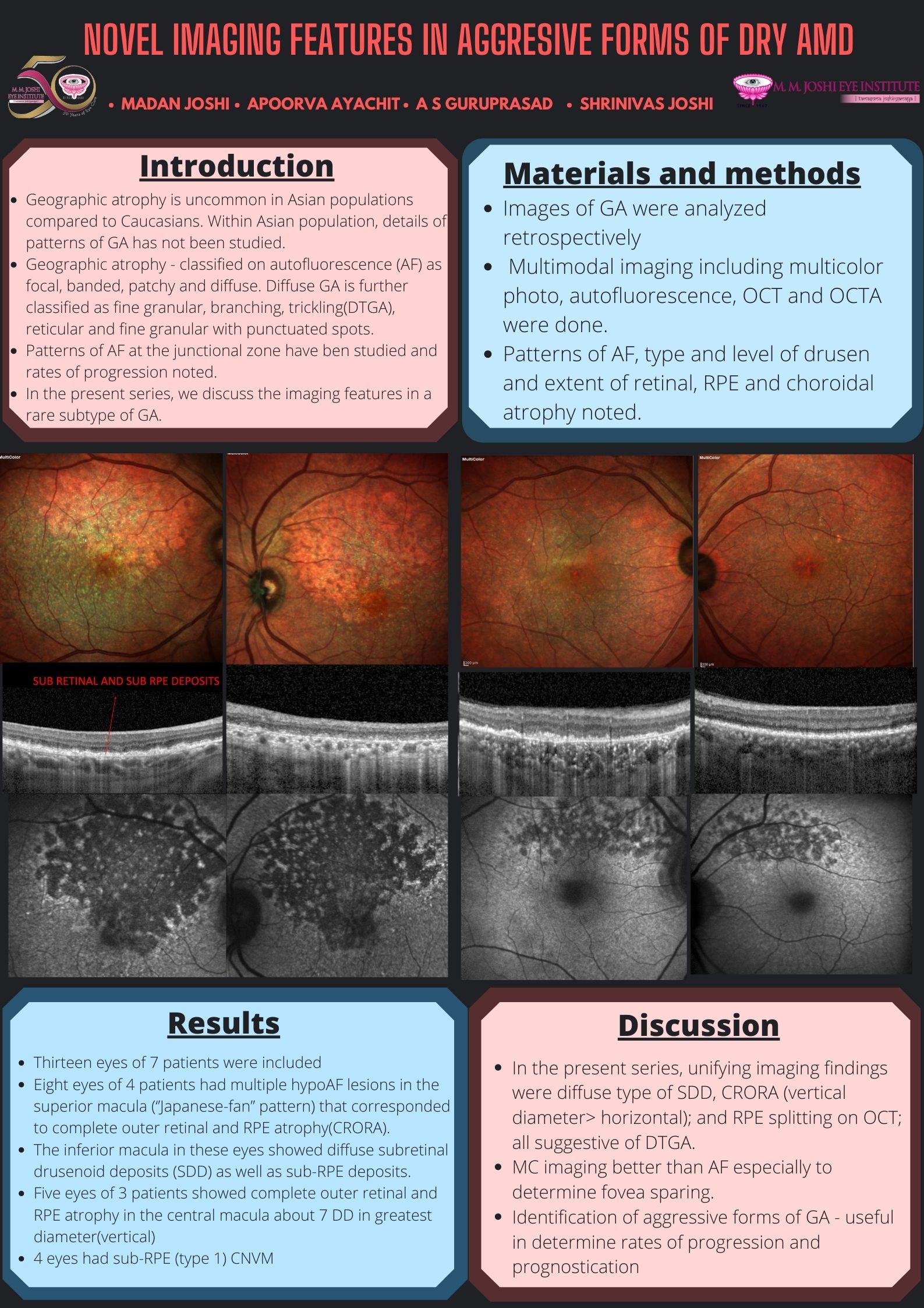
Thirteen eyes of 7 patients were included in this retrospective case series. Mean age was 68.23+/-13.14 years. F:M ::4:3. All eyes underwent multimodal imaging including multicolor, AF, OCT and OCTA. Eight eyes of 4 patients had multiple hypoAF lesions in the superior macula (‘’Japanese-fan’’ pattern) that corresponded to complete outer retinal and RPE atrophy(CRORA). The inferior macula in these eyes showed diffuse subretinal drusenoid deposits (SDD) as well as sub-RPE deposits. Five eyes of 3 patients showed complete outer retinal and RPE atrophy in the central macula about 7 DD in greatest diameter(vertical). 4 eyes had sub-RPE (type 1) CNVM. The unifying imaging findings were diffuse type of SDD, CRORA (vertical diameter> horizontal); a distinct ‘’Japanese fan’’ pattern atrophy and RPE splitting; all suggestive of diffuse trickling subtype of geographic atrophy. Identification of DTS- GA enables clinicians prognosticate in terms of progression and degree of vision loss.
Leave a Comment