
Dr.ANKITA GOEL
Dr. EVA RANI TIRKEY, Dr. SHASHI AGARWAL, PROF. PANKAJ CHOUDHARY
Abstract
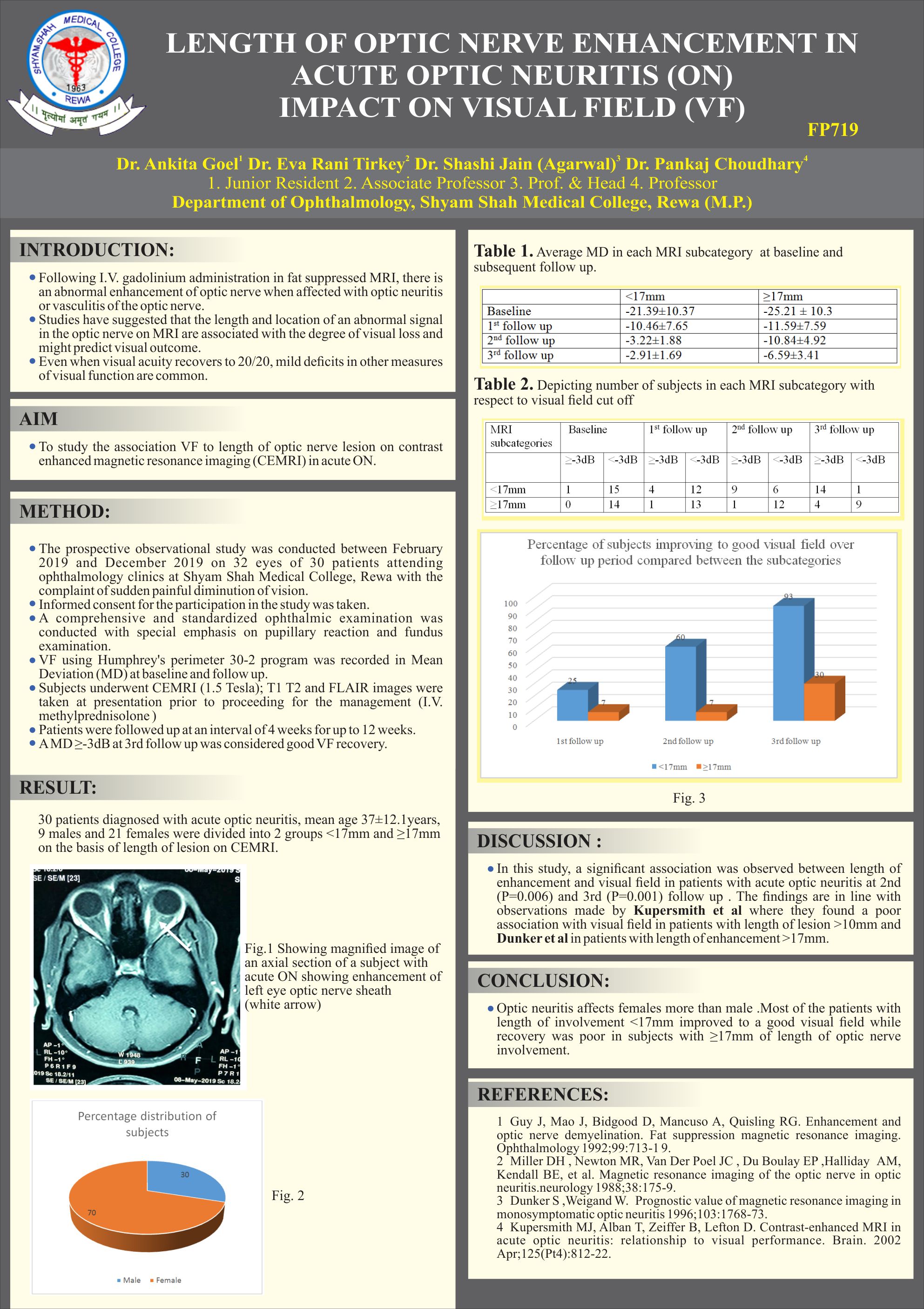
AIM: To study the association VF to length of optic nerve lesion on contrast enhanced magnetic resonance imaging (CEMRI) in acute ON.
METHOD: A comprehensive and standardized ophthalmic examination was conducted on 32 eyes clinically diagnosed as acute ON. They underwent CEMRI (1.5 Tesla) at presentation, were then treated with intravenous and oral steroid and followed up for 12 weeks. VF using Humphrey’s perimeter 30-2 program was recorded in Mean Deviation (MD) at baseline and follow up. A MD ≤-3dB was considered good VF.
RESULTS: 30 patients of acute optic neuritis, mean age 37±12.1years, 9 males and 21 females were divided into 2 groups <17mm and ≥17mm on the basis of length of lesion on CEMRI. 53% patients with <17mm involvement had a MD -2.9±1.64dB & 47% of ≥17mm had a mean MD -7.4±4.51 dB which was significantly (p=.001) less than patients in other group. CONCLUSION: There is significant association between length of lesion and VF loss.
Leave a Comment